Radiologist-led Care
촬영한 영상을 읽는 과정이
진단의 방향을 정합니다
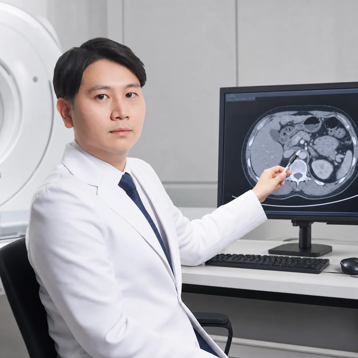
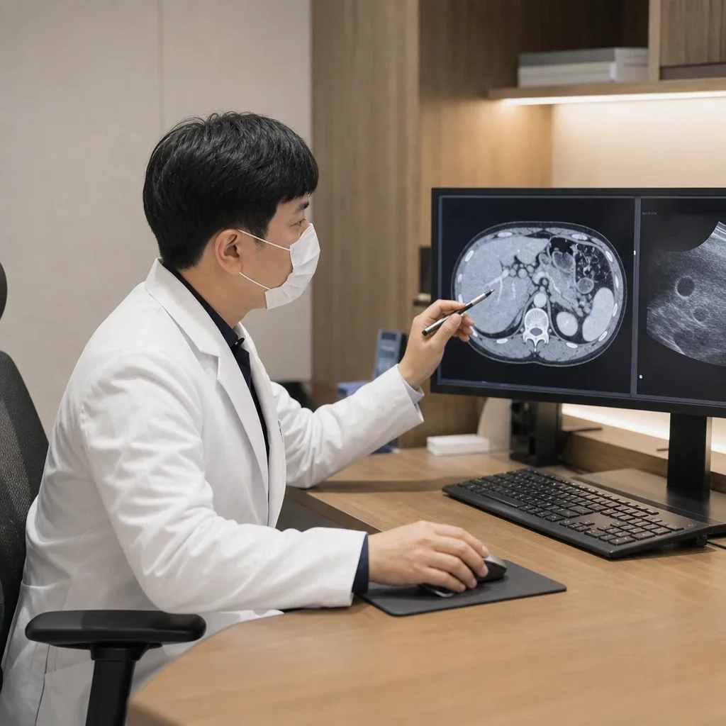
CT·MRI·X-ray·초음파는 검사를 시행하는 것만으로 끝나지 않습니다. 임상 정보와 이전 검사를 함께 살피고, 영상에서 확인되는 소견을 현재 증상과 연결해 해석해야 합니다.
박세진 대표원장은 서울대학교병원과 서울시립보라매병원 영상의학과 진료교수로 근무했으며 , 복부 영상의학 분야의 진료와 연구 경험을 바탕으로 검사를 판독합니다.
검사 전 판단부터
결과 설명까지 연결합니다
영상검사는 검사 종류와 촬영 범위를 적절히 선택하는 것부터 시작합니다. 의료진이 필요한 정보를 공유하고, 판독 결과가 다음 진료 계획으로 이어지도록 구성합니다.
증상과 이전 검사 확인
현재 증상, 진료 목적, 과거 영상과 검사 결과를 확인합니다.
검사 방법 선택
확인할 장기와 질환 가능성에 따라 CT·MRI·초음파·X-ray를 선택합니다.
영상의학과 전문의 판독
촬영 영상을 구조적으로 검토하고 필요한 소견을 판독문에 정리합니다.
진료과 협진과 결과 상담
필요하면 내과 의료진과 결과를 공유하고 후속 검사나 진료 방향을 안내합니다.
필요한 정보를 얻기 위해
검사 방법을 구분합니다
장기와 의심 질환에 따라 필요한 영상 정보가 다릅니다. 진료 후 적합한 검사와 촬영 범위를 안내합니다.
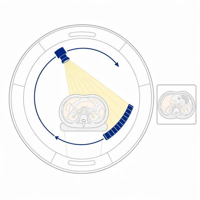
CT
흉부·복부·뇌·심혈관 등 장기의 단면 영상을 빠르게 확인합니다.
검사 상세 보기 → 02 · MAGNETIC RESONANCE IMAGING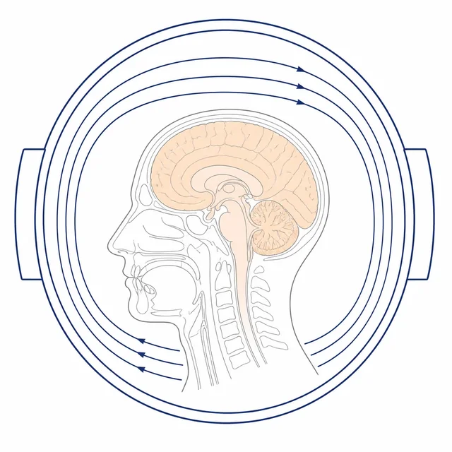
MRI
뇌와 복부 장기, 연부조직을 다양한 영상 기법으로 평가합니다.
검사 상세 보기 → 03 · ULTRASONOGRAPHY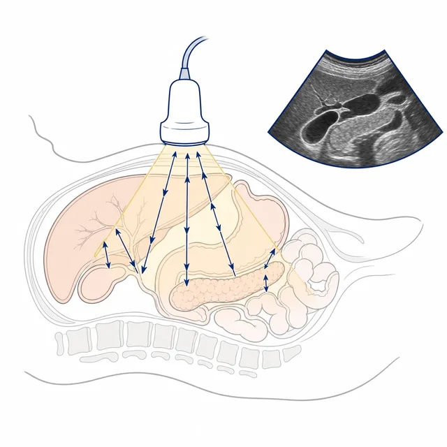
초음파
복부·갑상선·유방·혈관 등을 실시간으로 관찰합니다.
검사 상세 보기 → 04 · DIGITAL RADIOGRAPHY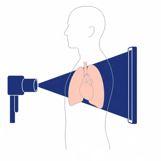
X-ray
흉부와 뼈를 포함한 기본 구조를 짧은 시간에 확인합니다.
검사 상세 보기 → 05 · BREAST IMAGING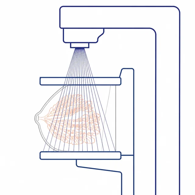
유방 영상검사
유방촬영과 초음파를 증상과 검진 목적에 맞춰 시행합니다.
검사 상세 보기 → 06 · THYROID IMAGING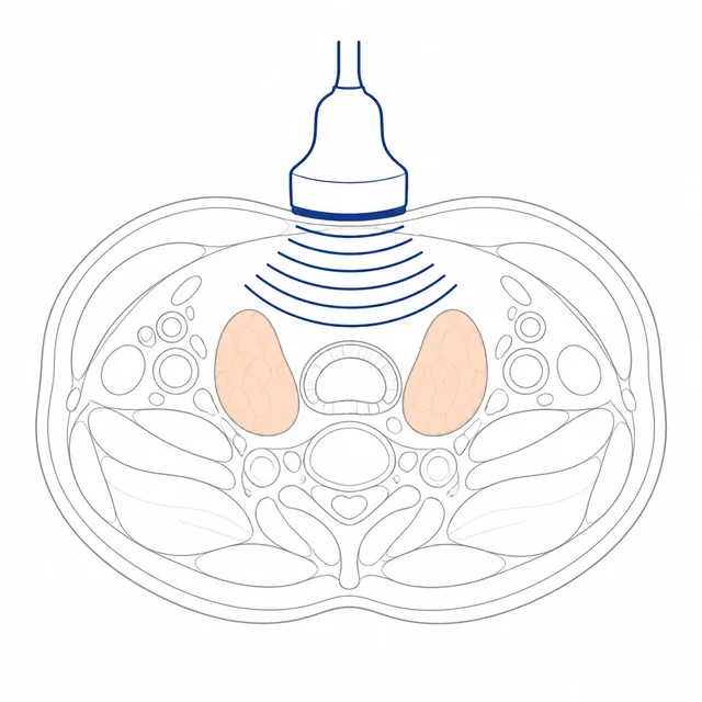
갑상선 초음파
갑상선 결절과 주변 경부 구조를 확인합니다.
검사 상세 보기 →Radiology Director
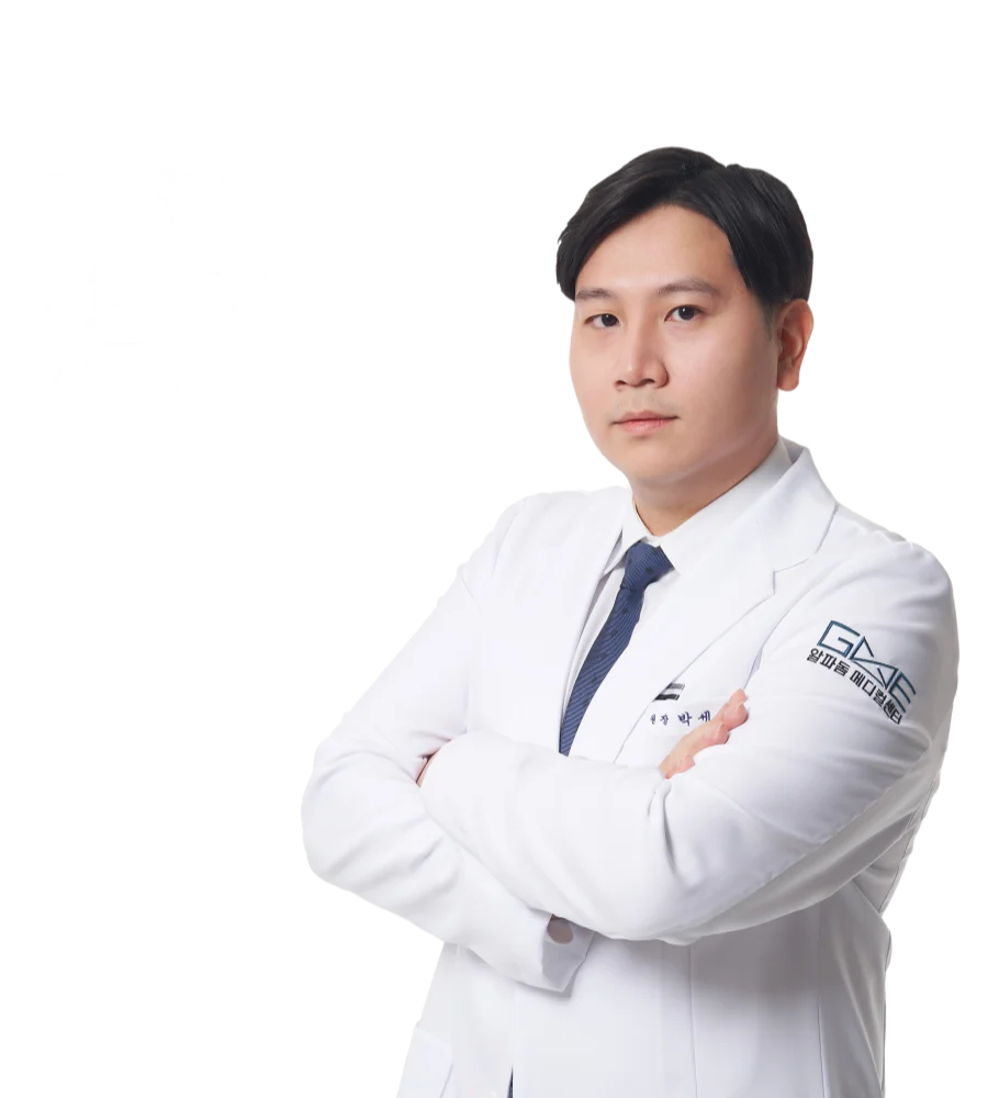
박세진 대표원장
영상의학과 전문의
서울대학교병원과 서울시립보라매병원 영상의학과 진료교수로 근무했습니다. 복부 영상의학을 중심으로 CT·MRI·초음파 판독과 영상 유도 치료 분야의 연구를 수행했습니다.
학력·경력과
연구 활동
영상의학 분야의 교육과 임상 경험, 학술 활동을 바탕으로 영상을 판독합니다.
학력·수련
- 서울대학교 의학대학 의학과 학사·석사·박사
- 서울대학교병원 인턴 수료
- 서울대학교병원 영상의학과 전공의 수료
- 서울대학교병원 영상의학과 전임의 수료
주요 경력
- 前 서울대학교병원 영상의학과 진료교수
- 前 서울시립보라매병원 영상의학과 진료교수
- 지앤이 알파돔 메디컬센터 대표원장
학회 활동
- 대한영상의학회·대한복부영상의학회 정회원
- 대한초음파의학회·대한ITA영상의학회 정회원
- 유럽복부영상의학회(ESGAR) 정회원
- 북미영상의학회(RSNA) 정회원
2021년 발표 논문
- Predictors of conversion surgery in patients with pancreatic cancer who underwent neoadjuvant or palliative FOLFIRINOX treatment using baseline and follow-up CT Abdom Radiol (NY). 2021;46(10):4765-4778.
- Preoperative assessment of the resectability of pancreatic ductal adenocarcinoma on CT according to the NCCN Guidelines focusing on SMA/SMV branch invasion Eur Radiol. 2021;31(9):6889-6897.
- Tumor Stiffness Measurements on MR Elastography for Single Nodular Hepatocellular Carcinomas Can Predict Tumor Recurrence After Hepatic Resection J Magn Reson Imaging. 2021;53(2):587-596.
- Reducing Pain by Artificial Ascites Infusion During Radiofrequency Ablation for Subcapsular Hepatocellular Carcinoma Cardiovasc Intervent Radiol. 2021.
- Switching Monopolar No-Touch Radiofrequency Ablation Using Octopus Electrodes for Small Hepatocellular Carcinoma: A Randomized Clinical Trial Liver Cancer. DOI: 10.1159/000512338.
- Virtual noncontrast images derived from dual-energy CT for assessment of hepatic steatosis in living liver donors Eur J Radiol. 2021;139:109687.
- LI-RADS v2018: how to appropriately use ancillary features in category adjustment on gadoxetic acid-enhanced MRI Eur Radiol. 2021.
- CT for lymph node staging of Colon cancer: not only size but also location and number of lymph node count Abdom Radiol (NY). 2021;46(9):4096-4105.
- Quantitative Ultrasound Radiofrequency Data Analysis for the Assessment of Hepatic Steatosis in Nonalcoholic Fatty Liver Disease Korean J Radiol. 2021;22(7):1077-1086.
2019~2020년 발표 논문
- Important CT and histopathological findings for recurrence and overall survival in patients with pancreatic ductal adenocarcinoma after neoadjuvant FOLFIRINOX Eur Radiol. 2020.
- Immediate Mild Reactions to CT with Iodinated Contrast Media: Strategy of Contrast Media Readministration without Corticosteroids Radiology. 2020.
- Prediction of microvascular invasion of hepatocellular carcinoma: value of volumetric iodine quantification using preoperative dual-energy CT Cancer Imaging. 2020;20(1):60.
- Deep learning-based decision support system for the diagnosis of neoplastic gallbladder polyps on ultrasonography Sci Rep. 2020;10(1):7700.
- Deep Learning for Chest Radiograph Diagnosis in the Emergency Department Radiology. 2019;293(3):573-580.
- Preventive Effect of Changing Contrast Media in Patients With A Prior Mild Immediate Hypersensitivity Reaction to Gadolinium-Based Contrast Agent Invest Radiol. 2019;54(10):633-637.
2017~2018년 발표 논문 및 주요 연구
- No-Touch Radiofrequency Ablation of VX2 Hepatic Tumors In Vivo in Rabbits: A Proof of Concept Study Korean J Radiol. 2018;19(6):1099-1109.
- Magnetic resonance elastography of healthy livers at 3.0 T Eur J Radiol. 2018;107:46-53.
- Prospective Validation of Intra- and Interobserver Reproducibility of a New Point Shear Wave Elastographic Technique Korean J Radiol. 2017;18(6):926-935.
- The prevalence, risk factors, and clinical outcome of balloon rupture in balloon-occluded retrograde transvenous obliteration of gastric varices J Vasc Interv Radiol. 2010;21(4):503-507.
검사 목적을 확인하고
결과까지 안내합니다
검사 종류와 조영제 사용 여부에 따라 준비 과정은 달라질 수 있습니다.
진료·문진
증상과 과거력, 이전 검사 자료를 확인합니다.
검사 계획
검사 부위와 촬영 방법, 조영제 필요 여부를 검토합니다.
촬영·판독
검사를 시행한 뒤 영상의학과 전문의가 영상을 판독합니다.
결과 상담
판독 결과를 설명하고 필요하면 후속 진료로 연결합니다.